This is restricted due to surgical images.
Surgical Delay for Extending the DIEP
Presentation by Dr. Joshua L. Levine
Transcription of above presentation:
Surgical Delay for Extending the DIEP | Introduction
Good afternoon. My name is Josh Levine, I’m speaking to you from New York City, and I’d like to thank Aldona for inviting me to share our recent experience with surgical delay for extending a DIEP. The surgical delay phenomenon is something that is a very well-known phenomenon in the surgical world. It’s an instrument by which we are able to improve the blood supply to any donor tissue.
We’ve been using this phenomenon recently to improve our ability to use the abdomen as a donor site in perforator flap breast reconstruction more effectively. We started over a year ago delaying the abdominal donor site in patients who have had previous abdominal surgery and likely weren’t thought of as candidates for the use of the abdomen in perforator flap breast reconstruction. We found that we could convert these patients to viable candidates for the use of the abdomen, and then subsequently, we started offering the delay to patients who just would benefit from having more abdominal tissue for breast reconstruction.
Surgical Delay for Extending the DIEP | A Well-Known Phenomenon
The delay is a very well-known phenomenon and it’s very well-described. Underneath the skin, there are a number of choke vessels separating one territory from another, and if the vessels that supply blood to one territory are transected as you see in this representation, the choke vessels will then open up so that the adjacent vessel then becomes responsible for supplying an area of tissue that it previously would not have been responsible for. So, the goal of the delay is to increase the amount of blood supplied to a tissue and the area of tissue by opening choke vessels in a very systemic way according to the vessels that we select, the vessels that we select being the perforator to preserve and the surrounding vessels are transected. Over the course of time, namely about one week or more, the blood vessel that has been selected will “learn” to perfuse the adjacent tissue and, therefore, increase the amount of tissue that one blood vessel, namely, a perforator, can supply.
Surgical Delay for Extending the DIEP | Patient Example 1
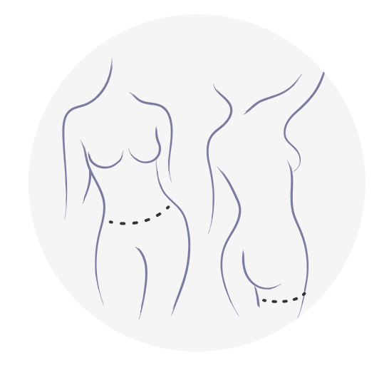
This is a typical patient who would like to use autologous tissue for breast reconstruction but due to an inadequate volume would never be thought to be a candidate. So, after getting an MR angiogram and identifying key perforators on either side of the abdomen, we go in preemptively, one week prior to the flap transfer, and we find the perforator that we’d like to preserve. This is the left hemiabdomen looking down from above the belly button, and here on the right we see the right hemiabdominal perforator, and each one of these perforators is preserved and everything else is transected except for a lateral skin bridge. So, we’ve taken out the blood supply from above and below and the deep circumflex and the superficial and all of the adjacent abdominal perforators. Only a skin bridge is left laterally, and the one perforator on either side of the abdomen that we will be using to transfer the flap. Then, subsequently in the operating room, we can use Doppler to follow the progress of the opening up of the choke vessels and the improve of perfusion in lateral tissue, and it’s very gratifying to see that a very medially based perforator after even a day or two, and certainly after a few days, will supply all the way out to the periphery even beyond zone 2 such that we are significantly increasing the amount of volume that one abdominal perforator can supply. Then, when we go to the operating room at least one week later, we can extend the design of the abdominal flap way beyond zone 2, even into a lateral hip area and towards the lumbar area, and once this tissue is harvested we can see that it perfuses very, very well.
Here’s the left hemiabdomen being harvested. We’ve opened the rectus sheath and as you can see, the perforator is quite nicely dilated, and once the flaps are harvested in situ we can see with the Doppler we can trace that signal all the way out beyond zone 2, and we can see that that tissue perfuses quite nicely in situ. The flap is harvested, is brought to the chest, the anastomoses are performed and then the flap is inset, and in this way we’ve achieved a much larger amount of volume than we could certainly have gotten with an ordinary perforator flap.
Here’s another example of a patient that had significant abdominal morbidity from repeated liposuction for salvage of failing implants. She developed chronic pain and scar tissue and even some significant encapsulated seromas, which are represented here in this MR angiogram by this scar tissue that you see. This is an encapsulated seroma.
This was an abdomen that I think most of us would probably not have considered for use in perforator flap breast reconstruction due to the extensive scarring, and in fact, when the abdomen was explored, this large encapsulated seroma cavity was excised along with a few others, but in spite of that there were some viable perforators there and they were preserved, one on either side, and again, leaving a skin bridge laterally as you can see here.
So, after waiting a week, and here again the lateral skin bridge on the patient’s left hemiabdomen [00:07:50 and] the belly button being here, the patient was taken back to the operating room and, again, the perforators are readily identified. There are no other blood vessels in this area. So, they’re easy to find and quite nicely dilated as you can see. So, the rectus sheath is open, the perforators are dissected in the usual manner, and the flaps are then harvested based on those perforators. Here she is postoperatively, nice autologous reconstruction in a patient who really would not have been a candidate for the use of the abdomen.
Surgical Delay for Extending the DIEP | Patient Example 2
Here’s another example of what we typically see, which is a glistening seroma cavity which is sort of scarred off the perforator, and in order to do these dissections we found that it’s very useful to actually enter the rectus sheath medial to the perforator because it’s really surrounded by scar tissue in many cases. And so, we can approach this very cautiously and then do the dissection under direct division.
Surgical Delay for Extending the DIEP | Patient Example 3
Here’s another example of a patient who had implant failure, chronic pain, dissatisfaction with implant reconstruction. You can see an old scar here on her right hemiabdomen. This is a scar from an excision of a dermoid tumor that she had when she was a child, and ordinarily we wouldn’t think that that would be much of a problem, but of course MR angiogram is essential in the planning of these surgeries and, as you can see in this right hemiabdomen, an absence of the rectus abdominis and a complete transection of the DIEP, which is readily available on her left side. So, this patient really was not a DIEP candidate on the right side, and we took her to the operating room hoping to perform a delay of the superficial vessels on the right side.
Interestingly though, she did not have a superficial on the right, and so we ended up doing a first-ever delay of deep circumflex on the patient’s right-hand side. The deep circumflex is a vessel that we were previously using quite frequently in an operation that we call the SHaEP flap or stacked hemiabdominal extended perforator flap. And so, it’s a vessel that is very reliable, but it’s quite small. And so, to use it in conjunction with a DIEP and connect into the DIEP I think is something that works very, very well, but to use it as the only vessel for an abdominal perforator is somewhat risky without the delay. However, the delay in this case proved to be quite effective. We can hear the vessel over the deep circumflex after the delay, which is very gratifying in the office postoperatively.
And then, here is the intraoperative picture of the deep circumflex after the delay. This is the right hemiabdomen. The deep circumflex is nicely dilated, and it does indeed very nicely perfuse the right side. So the right side was a deep-circumflex first-of-its-kind delay, and the left was a DIEP-1 which we’ve had much more experience with – done about 30 flaps of a delayed DIEP.
And then, we were able to confirm nice perfusion of both hemiabdominal flaps. Here’s the right side, the deep circumflex very, very nicely perfused, and then of course the DIEP on the left.
So, once the flaps are isolated, they have transferred, and again, a huge increase in the amount of volume that we can get.
Surgical Delay for Extending the DIEP | Example 4
Here are a couple of examples. This is a patient who had bilateral mastectomy with a delayed DIEP flap breast reconstruction. Here’s another patient – typical patient who had extensive abdominal liposuction and would not have been considered a candidate, and in fact on the MR angiogram you can really see the extent to which the abdominal liposuction creates scar tissue and, of course, decreases the volume of harvestable tissue, and this patient was converted into an abdominal candidate after a miserable time with her implant reconstruction.
Surgical Delay for Extending the DIEP | Example 5
Here’s another example of a patient with inadequate abdominal volume for two breasts.
So, we’ve been using this technique now to create candidates out of patients who wouldn’t have been candidates to improve the candidacy of patients who might have had inadequate tissue volume from the abdomen. We’ve done it now on a number of cases on the DIEP, a few in the SIEA, and we’ve done one DCIA. It really does change the game in terms of using the patient’s abdomen, and I think this will be a huge improvement in our ability to offer abdominal-based perforator flap breast reconstruction to many, many more women.
We’re still gaining experience and learning from our experience. We’ll be publishing it soon in PRS. But, the bottom line is that we’ve done a lot of work with stacked or multiple-flap procedures, and what this does for us is it enables us to use one donor site as opposed to multiple, one pedicle as opposed to multiple, and it dramatically simplifies the operation and really gives you an enormously powerful tool in perforator flap breast reconstruction from the abdomen.
So, that is the conclusion, and I thank you very much once again for allowing me to share this information. I look forward to talking with everyone in the panel discussion. Thank you again.


