Breast reconstruction education
What is Autologous Breast Reconstruction?
Autologous breast reconstruction rebuilds the breast using a woman’s own tissue instead of an implant. “Autologous” means “from the same person.” Skin and fat are taken from an area of the body with extra tissue and shaped into a new breast. With microsurgery, the tissue’s blood vessels are reconnected in the chest, giving the reconstructed breast its own living blood supply.

The Three Types of Autologous Breast Reconstruction
There are three main ways a woman’s own tissue can be used to rebuild the breast:
Type 1
Perforator Flaps – True Muscle-Sparing Techniques
A perforator flap is the most advanced form of autologous breast reconstruction. Only the tissue needed to rebuild the breast is taken from the donor site: skin, fat, and the small blood vessel that supplies them, called a perforator. The underlying muscle is left completely intact. This sets perforator flaps apart from older muscle-based flaps like the TRAM.
For that tissue to survive in its new location, it needs a blood supply. This is where microsurgery comes in:
Each perforator flap is named for the blood vessel it uses, and the “P” in each acronym stands for perforator. The most common is the DIEP flap, which uses extra skin and fat from the lower abdomen.
Skin, fat and the perforator blood vessel. The muscle stays in place.
The tissue and its blood vessel are fully detached and shaped into a new breast.
Under a microscope, Dr. Levine reconnects the vessel to blood vessels in the chest.
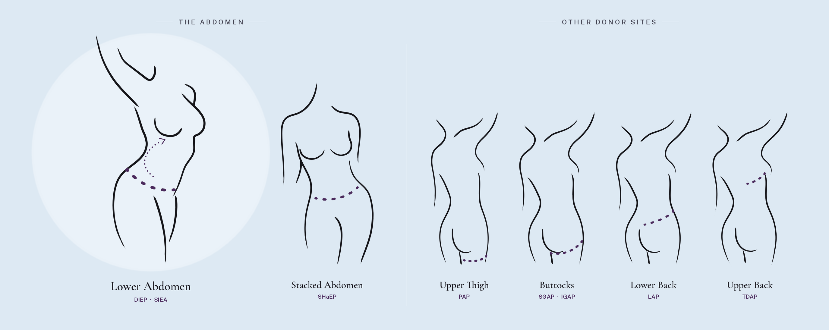
Common Donor Sites
The donor site depends on each woman's body type, where she carries extra tissue, and how much volume is needed.
Lower abdomen
The most common donor site, using the same tissue removed in a tummy tuck.
Abdomen and flanks
A stacked flap that combines tissue from both sides of the abdomen into a single breast. Ideal for women with less abdominal tissue.
Back of the upper thigh
Tissue from the upper inner and back thigh, with the scar hidden in the buttock crease.
Buttocks
Tissue from the upper or lower buttock, for women with more volume there than in the abdomen.
Lower back and flanks
Tissue from the "love handle" area above the buttocks.
Upper back
Tissue from the back below the armpit, often used for partial reconstruction.
Type 2
Myocutaneous Flaps – Muscle-Sacrificing
A myocutaneous flap moves skin, fat and muscle together. The muscle is taken because it contains the blood vessels that keep the transferred tissue alive. The most common myocutaneous flap is the TRAM. Other examples include the latissimus dorsi flap and the TUG flap.
TRAM Flap
Uses skin, fat and the rectus abdominis muscle from the lower abdomen to rebuild the breast. It can be done two ways. In a pedicled flap, the tissue stays attached to the muscle and is tunneled up to the chest. In a free flap, the tissue is fully detached and its blood vessels are reconnected in the chest.
Either way, the abdominal muscle is lost, which can weaken the core and raise the risk of a bulge or hernia.
Latissimus Dorsi Flap
The latissimus dorsi is a large muscle in the back. It is most often used together with an implant. The muscle is brought around from the back to the chest, where it covers the implant and adds soft tissue that thin or radiated skin may need.
For some women, there is enough skin and fat on the back to rebuild the breast with the latissimus alone, without an implant.
Type 3
Fat Grafting
Fat grafting is a technique in which fat is harvested from under the skin using liposuction. The fat is then processed and injected back into the chest to enhance or improve a reconstructed breast.
In some cases the entire breast can be reconstructed with the patient’s own fat. This requires preparing the chest skin and muscle with an expansion device called BRAVA, and the process takes many months and several operations. BRAVA expansion and fat grafting can also be used to improve or enhance an already reconstructed breast.
Unlike flap reconstruction, fat grafting does not transfer tissue with its own blood supply. Only a portion of the injected fat survives, which is why multiple sessions are usually needed and why it is most often used to refine or add modest volume rather than rebuild the entire breast.
Autologous Reconstruction vs. Implants
Because an autologous reconstruction is living tissue, it looks, feels and ages like a natural breast. It is warm and soft, and it changes with the body as weight goes up or down. There is no device that can rupture, harden from capsular contracture, or need replacing years later.
The tradeoff is a longer initial surgery and recovery, since there is a donor site as well as the breast. For most women, the result is a single, lasting reconstruction instead of a lifetime of implant maintenance.
| Comparison | Your own tissue | Implants |
|---|---|---|
| Look and feel | Warm, soft, living tissue | A device beneath the skin |
| Over time | Ages and changes with the body, including weight | Does not change with the body |
| Device risks | None: no device to rupture or harden | Rupture, capsular contracture |
| Future surgery | Typically a single, lasting reconstruction | May need replacing years later |
| Initial surgery | Longer surgery and recovery, with a donor site | Shorter initial surgery |
Pioneering natural reconstruction
Meet Dr. Joshua L. Levine
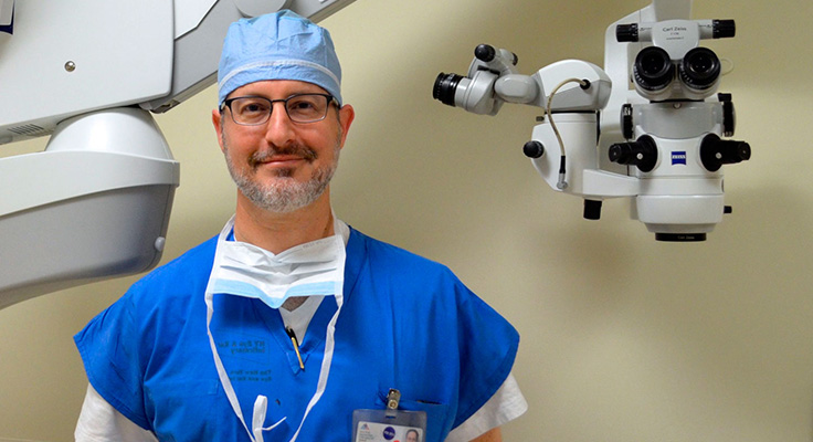
Dr. Levine performs the full range of perforator flap procedures, including DIEP, SIEA, SHaEP, PAP, GAP, LAP and TDAP flaps. He has further advanced this field, developing first-ever techniques in extension flap and stacked flap procedures.
These pioneering techniques now mean that all women can restore the look and feel of their own warm, natural breasts regardless of their body type.