This is restricted due to surgical images.
Breast Reconstruction Overview and Perforator Flaps
Presentation by Dr. Joshua L. Levine
Transcription of above presentation:
Breast Reconstruction Overview and Perforator Flaps | Intro
Hello, I’m Dr. Joshua Levine in New York City. Today I’m going to be going over some basic concepts of breast reconstruction and an overview of perforator flaps.
This is a talk that I gave back in 2015 at a FORCE meeting in order to educate patients who were dealing with the risk of having breast cancer. FORCE stands for Facing Our Risk of Cancer Empowered, and it’s an organization that is geared towards providing information for patients who have genetic mutations which predispose them to a high risk of breast or ovarian cancer. The reason that I’m reviewing this topic is because it really is a great overview to give you a basic understanding of what breast reconstruction is all about and to learn some of the options.
This is a patient who has had bilateral prophylactic mastectomy and breast reconstruction, and this is the type of patient that we’re going to be talking about. This is a patient who is at high risk for breast cancer. She elected to have both of her breasts removed without having cancer, and that’s called prophylactic mastectomy. She had nipple-sparing mastectomy and she had autologous breast reconstruction. These are some of the concepts and terms that I’ll be explaining in this short video.
Breast Reconstruction | Then and Now
Let’s just talk a little bit about breast reconstruction. We’re talking about replacing a normal part of the female anatomy after a mastectomy. Mastectomy can be done for cancer or it can be done for a high risk of developing breast cancer in order to prevent cancer, and in that case it’s called prophylactic mastectomy. When the breast is removed, it can be replaced with either implants or the patient’s own body tissue, and when we use the patient’s own body tissue we call that autologous breast reconstruction.
Now, the old way of replacing the breast with a patient’s autologous tissue was to use muscle and skin and fat. We call these myocutaneous flaps or muscle/skin flaps. The reason that we used to do it this way is because a lot of the blood that supplies the tissue that we want to transfer goes through the muscle. So, the thinking was that it was necessary to take the muscle along with the skin and fat, but we know now that that’s not true. We can take only the tissue that we want to take to make the breast as long as we take along with it the key blood vessels that supply it. Those blood vessels are called perforators, and so the surgery or the technique that we’re talking about here has been perforator flaps and that refers to the tissue that’s transferred along with its perforating blood vessel.
Now, what does the perforating blood vessel perforate? It perforates a muscle, and just to go back to that concept for just a minute, prior to the advent of the perforator flap it was thought that the muscle had to be taken with the skin and fat. Again, those were called muscle/skin flaps or myocutaneous, and you might have heard them referred to in some of the more common operations that are done with that technique, namely, the TRAM flap or the latissimus flap. Now, both those operations refer to harvesting tissue from the patient’s body but taking along with it a very important muscle. Perforator flap does not do that. The perforator flap takes only the skin and the fat that we need to make our new breast and the key perforating blood vessel that supplies it.
Breast Reconstruction | Perforator Flap Donor Site
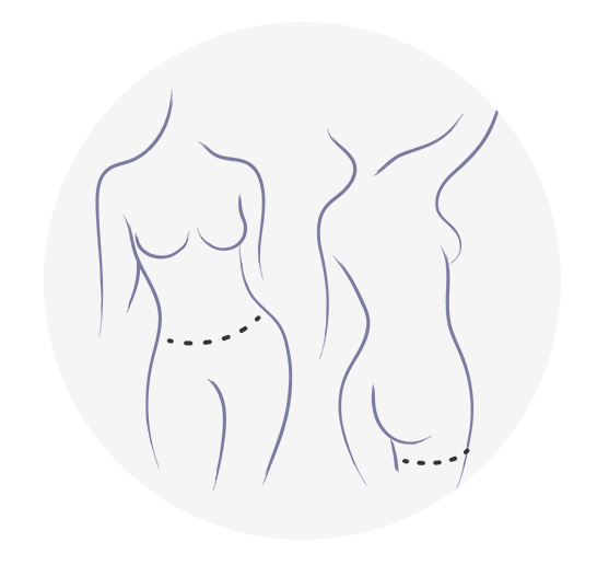
Now, we typically use the abdomen as our primary donor site because in many women who are of breast-cancer-related-issues age they may have a little extra in the abdomen to donate, particularly if they’ve carried pregnancies in the past. So, the abdomen is a great donor site for breast reconstruction whenever it’s available, but there are other donor sites, and some women may have had too much surgery on the abdomen to make it a viable option. Some women may be just too thin and there might not be enough tissue there to harvest to make one or two breasts, and in those cases we can look elsewhere and, typically, we’ll look for other areas where there is extra fat, namely, the thigh or the buttocks.
All of these areas can be donor sites, and they’re all donor sites with names. When we take it from the abdomen, we call it the DIEP or the SIEA; when we take it from the buttock, the SGAP or the IGAP; and the thigh, the PAP or the TUG flap. The names aren’t that important, but what you do need to remember is that these are perforator flaps, and if it has a P in it it’s a perforator flap because the P stands for perforator.
Now, many patients ask, “Is this a new operation?” and the answer is no. When we first started doing these operations 20 years ago, of course they were quite new, but they’re so ubiquitous at this point in time that it’s actually required that a nationally certified breast cancer treatment center perforator flaps in order to be certified. So, this has become a standard part of breast reconstruction.
Breast Reconstruction | Replacing the TRAM Flap
Now, just to digress a moment to give you some information about anatomy which will help you to understand the perforator flap, let me tell you what a TRAM flap is. The TRAM is a transverse rectus abdominis muscle flap or myocutaneous flap, and the way this works is this tissue that we want to use to make the breast is taken from the lower abdomen, and while it’s still attached to the muscle, namely, the rectus abdominis, it is swung around into the chest in order to make a breast.
Now, the reason that it’s swung around and still attached is because the blood vessels that supply that tissue that we want—the skin and fat of the lower abdomen—travel within the rectus abdominis. In fact, they perforate through it. So, once we figured out that the perforator blood vessels could be taken out of the muscle without damaging or sacrificing the muscle, the perforator flap has [00:07:41 largely] replaced the TRAM flap. The reason this was so important is because the TRAM flap does in fact sacrifice a major muscle in the abdomen, and that’s when you’re using one side, but men and women these days undergo bilateral or both-sides mastectomy and to take both muscles is a significant price to pay. It can cause a hernia, abdominal wall weakness and a significant amount of postoperative pain and morbidity.
The blood vessels that supply this tissue run through and also underneath the rectus abdominis. They come from above but they also come from below, and when they come from below they’re coming off of a blood vessel called the deep inferior epigastric, and that’s where you get the name for the DIEP. It stands for deep inferior epigastric, and the P of course, again, is our perforator.
Breast Reconstruction | MR Angiography for Imaging Perforator Flaps
Now, when we go in to harvest one of these flaps, we know exactly where we’re going, and that’s because we have developed a protocol for MR angiography. You might have heard of the MRI, which is done very frequently to make diagnoses. We can also use MR—which is magnetic resonance—technology and do an angiogram or look at the blood vessels, and what we’re looking at here is an MR angiogram in a technique that my team developed back in 2006 and we still use on every case to this day.
What you’re looking at is a cross section through a patient’s abdomen. You can see at the very top the skin and underneath that the fat, and underneath the fat you can see the two rectus abdominis muscles parallel to one another, like this, and coming right through one of those muscles you see a while line which represents a blood vessel. That is a perforator, and it has perforated through the rectus abdominis and it has come off of the deep inferior epigastric.
When we look at this in the operating room, we find the perforator. This represents an intraoperative diagram of what things look like once we get in there. You can see the belly button at the top of your screen, and then the blood vessels coming through the rectus abdominis and into the flap. The dissection has been done with the muscle fibers being very delicately and gently separated such that the muscle is still functional and preserved after this operation, and the perforator is taken along with the flap and the muscle is closed right back up. The tissue is then brought to the chest and plugged into a blood vessel in the chest, namely, the internal mammary artery and vein, and this is what it looks like in a diagram.
Breast Reconstruction | Supplying Enough Tissue for Perforator Flap Reconstruction
Many times, patients have had cancer, they’ve had bilateral or both-sides mastectomy, and in most women there is enough abdominal tissue to harvest to make two breasts. This is an example [00:11:25 thereof] with reconstructed breasts. Here’s another example of how the procedure can be a cosmetic advantage not only to the breasts, obviously, but to the abdomen as well. Here’s another example of a unilateral or on one side—the patient’s left side was reconstructed with her abdominal perforator flap or DIEP—and here’s another example of a bilateral.
Now, some women don’t have enough abdominal tissue or maybe they would just prefer not to use the abdomen, and in those cases we have other options. We can use the posterior thigh, which we call the PAP flap, and I have a video explaining the PAP flap also but here’s a brief overview. This is called the PAP flap because it comes off of the profunda artery. So, it’s a profunda artery perforator, comes from the back of the leg.
Breast Reconstruction | BRCA Patient
Here we have a very thin BRCA patient, which I showed you initially – very little abdominal volume but a little bit of extra in the thigh area. So, we can use her thighs, and here she is after surgery. From the front, it’s hard to tell she even had surgery. The incisions are lateral to the nipples and she had nipple-sparing mastectomy, and those are her scars on the backs of her legs. This is the area from which we took the tissue, the PAP flap. Here she is again after breast reconstruction.
This is a patient who could have had either a DIEP or PAP flaps. She’s had implant reconstruction, which didn’t work out very well as you can see, radiation on the left with significant deformity and capsular contracture, and she really is fed up with her implants and ready to have them removed, but she didn’t want a scar across the front of her abdomen. She did have adequate volume in the thigh area below the waist, and so this is what we elected to do for her. This is the design of the flap and the dots there that you see represent the perforators, which have been preselected with MR angiogram or MRA.
After the surgery, this is what she looks like. The implants have been removed and replaced with her thigh tissue, and she’s had reconstructed nipples with nipple and areola tattooing.
This is the before and after. You can see the terrible deformity that she had before and the nice natural breast reconstruction that she has after, and no disruption to the contour of her buttock and well-hidden scars below the buttocks there.
This is a postoperative picture of the scars on the backs of her legs on the PAP flaps, and here you see the same patient in profile.
So, in summary, I would say that knowing your options gives you power and authority to make the best decision for yourself. In my opinion, everyone is a candidate for autologous tissue. That means that if someone tells you that you cannot use your own body tissue and that you have to have implants I would very highly recommend that you seek a second opinion, and I think that knowledge will help you to make a decision that you’re ultimately very comfortable with.
Thank you so much for listening, and I encourage you to check out my other videos on similar topics. Thank you again.


