
PREOPERATIVE IMAGING (BEFORE SURGERY)
Our advances in pre-operative imaging have provided for a detailed plan or surgical “road map” that affords us a high level in the evaluation and technical planning that yields greater efficiency in true, muscle-sparing natural tissue breast reconstruction (perforator flaps.)
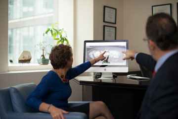
Back in 2005 when CT angiography was first being done preoperatively for perforator flaps, Dr. Levine immediately recognized the value of this information. Dr. Levine and his team with Dr. Prince of Weill Cornell Imaging and team developed the first protocol for obtaining the same valuable information without radiation through magnetic resonance angiogram (MRA.) Using their specific protocol with MRA is a way to see the tiny perforating blood vessels in performing the advanced microsurgical breast reconstruction with perforator flaps. The information we get from MRA provides a map of the best blood vessel that supplies tissue to the section of skin and fat “flap” transferred to recreate the breast.
Imaging results afford us the ability plan the surgery in fine detail — blood vessels are evaluated and selected in advance of surgery. This information saves time in the operating room and makes the operation much simpler and safer.
 INTRAOPERATIVE IMAGING (DURING SURGERY)

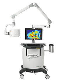
SPY Imaging is used during perforator flap surgery to evaluate and confirm perfusion, or adequate blood flow to the “flap.”