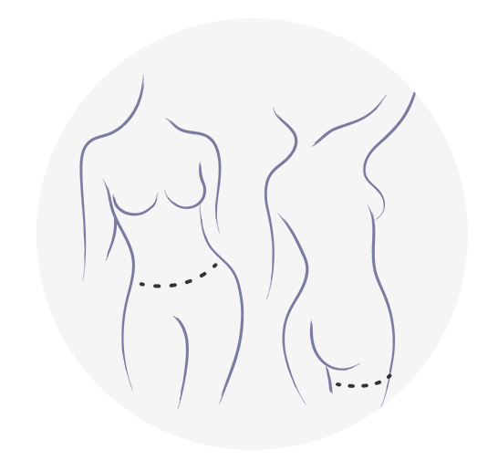
This is restricted due to surgical images.
More Is Better: Multiple and Delayed Flaps in Breast Reconstruction
Presentation by Dr. Joshua L. Levine
Transcription of above presentation:
Multiple and Delayed Flaps in Breast Reconstruction | Introduction
Hello, my name is Dr. Joshua Levine. I’m a microsurgeon in New York City, and in this video I’ll be describing some concepts related to autologous breast reconstruction or using a patient’s own body tissue to reconstruct breasts after mastectomy. I’ll be going into some advanced topics, but mostly I’ll be discussing the concept that more is better, meaning that for a plastic surgeon in order to reconstruct a cosmetically appealing and appropriately shaped and sized breast, the more volume of tissue that we have to deal with the better outcome we can provide and the more beautiful and aesthetic breast we can reconstruct.
The patients that I will be presenting here will all be patients who have had previous breast reconstruction with implants, and the implants have failed for one reason or another, which is very common, which we’ll discuss.
Just to give you an example of what we’re talking about when we talk about implant failure, I’ve been doing this for almost 18 years and I’ve seen a lot of implant-failure patients. That means that they’ve had reconstruction with implants after mastectomy and for whatever reason they can no longer tolerate the implant reconstruction. Over the years, I’ve kept an extensive list of some of the direct quotes that patients have mentioned in their consultation with me, and this is just a very, very brief list of the very extensive list of quotes that I have collected. And so, if you want to pause on this slide for a while and read through this, certainly if you’ve ever had implant reconstruction for breast reconstruction after mastectomy, you could probably find a few of these quotes that you can relate to, and if you’re contemplating implant reconstruction as opposed to autologous reconstruction it might be a good idea to familiarize yourself with some of these ideas.
Multiple and Delayed Flaps in Breast Reconstruction | Stacked DIEP
So, I’m going to jump right in here. The idea that more is better is something that we’ve known for a long time. This is a unilateral breast reconstruction, meaning one side – the patient’s left breast has been reconstructed. Actually, no. I’m fooled by the quality of the result. It’s actually her right breast that’s been reconstructed. The left breast has been lifted.
This was a thin patient who needed only one breast to be reconstructed, in other words, unilateral. And so, in order to provide a substantial amount of volume, we used both sides of her abdomen. When we divide the abdomen in halves, we call them hemiabdomen or hemiabdominal flaps, and in this case we used them both together and stacked them on top of each other, and we call that a stacked DIEP flap.
So, she had a stacked DIEP, and the idea was to get more volume. By volume, I mean just more tissue to work with.
This is what the double or stacked flap looks like after it’s harvested from the patient. This is the undersurface, the fat side. It’s the two halves of the abdomen. They’re still together. They’re contiguous. You can see the two blood vessels there that supply one side and then the other. When we shape it and bring the edges together, we can make a very reasonable shape and make a nice-looking breast out of it, and that’s the whole point of doing a stack and that’s the whole point of getting more volume to get more projection and more control over the result.
So, that’s a concept that we’re very familiar with, and we’ve done a lot of flap combining in order to achieve this in many different clinical scenarios. In other words, this is a list that is not even complete. This is a list of the flaps that we have combined together, and I know some of these terms are unfamiliar. The DIEP up at the top is probably familiar. But, all of these letters represent different autologous flaps that are taken from the patient’s body, and all of the different points that are made here are combinations of different flaps that have been used in order to provide more volume.
So, when you have a thin patient or an issue with getting enough volume, you have to get creative sometimes and there are a lot of different options for combining flaps. Again, the whole idea is that more volume is better because it gives you more options in terms of where you put the tissue, how you orient the tissue, and which tissue you use and which you don’t use.
Multiple and Delayed Flaps in Breast Reconstruction | Two Flaps is Better Than One
So, we’ve determined that two flaps is better than one, and by example I explained to you about the stacked DIEP for a unilateral breast reconstruction, and if we accept the fact that two is better than one, then we can probably agree that four is better than two. In other words, if you’re doing a bilateral, meaning both-breast reconstruction, and you need enough volume for two breasts and the abdomen is not enough, one common way that we address that is by stacking, again, different flaps. The DIEP can be placed on top to make the upper part of the breast, and a PAP flap or a profunda artery perforator can be used from the back of the leg to make the lower part of the breast, and this is a very common combination of flaps that we use when we are facing a volume deficit and perhaps a thin patient or a patient who just would prefer to have a larger breast after mastectomy.
Multiple and Delayed Flaps in Breast Reconstruction | Implant Failure Reconstruction #1
Here’s an example of a patient with implant failure. As I said, I was going to be discussing implant failure cases. This is a woman who had breast reconstruction after mastectomy for cancer. She had two implants placed to make new breasts and they were very, very unpleasant. She was in chronic pain and you could refer back to that first slide there – some of her quotes were certainly up there. But, as you can see, she’s a very thin woman and it would be very difficult to find one donor site to supply enough tissue for two breast reconstructions. So, you can see that we used her abdomen, and the postoperative picture on the right after her implants have been removed and replaced with her own body tissue, those are reconstructed breasts with reconstructed nipples on the right.
We used her abdomen, yes, but it wasn’t enough. And so, in order to provide the extra tissue volume that we needed, we also used the backs of her legs. This is a postop picture, pre- and postop, left and right, and on the right you see the scars from the PAP flaps. Let me just go back to that real quick. That would be right here, scars from the PAP flaps.
Multiple and Delayed Flaps in Breast Reconstruction | Implant Failure Reconstruction #2
Here’s another example, a patient who had implant failure, very miserable, with many, many years of failed implants that she just couldn’t live with anymore, and not a thin abdomen but certainly not enough to get volume for two breasts. And so, we ended up using her abdomen there. You can see her scar across the abdomen there. We also used her thighs. And so, you don’t see the scars on the thighs because they’re in the back.
Here’s another example. This is a young woman who had cancer, and she didn’t have implant failure, actually. She had a mastectomy and reconstruction and not enough volume for two breasts, so we used both the abdomen and the thighs. You can see here the scar from the mastectomy—so this postop after reconstruction—and here are the scars on the backs of the legs from the PAP flap.
So, these are some great examples of combination flaps, and in these cases we used the DIEP and the PAP.
Now, in order to maximize the amount of volume we could get from the abdomen, we learned how to do this operation, which we wrote about and published in 2018. We called it a stacked hemiabdominal extended perforator flap or the SHaEP flap, and what this allowed us to do is to acquire more volume from the abdomen, from each hemiabdomen, to make two breasts or bilateral breast reconstruction. The perforator that we use in a DIEP only supplies so much tissue volume, and so if we want to get more, in other words, take farther out towards the hip area, we have to do something creative, and in these cases we extended the design of the flap out towards the hip so that we have one vessel going into the main area of the DIEP. This would be in the medial portion near the belly button. And then, to get this lateral hip tissue, we would take this extra vessel and plug into it. So, this was an extra blood vessel. We had two blood vessels for each flap. The names of those blood vessels are the DIEP, again, in the medial portion—that’s the standard DIEP—and it’s hooked into a DCIA or deep circumflex iliac, just another vessel laterally. It doesn’t have to be the DCIA, and I’ll show you examples of other vessels that can be used, but that’s what we did in order to maximize the abdominal volume.
Multiple and Delayed Flaps in Breast Reconstruction | Using 3 Pedicles #1
Building on this concept that more is better, if two is better than one and four is better than two, then we can also see ourselves in situations where even more than two is better, and in this case we used three pedicles or three flaps for each breast reconstruction. So, this patient is preoperative, she’s got breast cancer, and the volume requirement is high. We’re only going to use each half of the abdomen for each breast, and if we did a standard DIEP we would get about expert much tissue – not enough. If we extended it out to here with the SHaEP flap and included this vessel, we might get enough, but if we come all the way around to the back we’ll certainly get enough.
But, in this case, it required a third blood vessel for each flap. So, this is just taking it to an extreme here. We go all the way around, and this is us turning the patient intraoperatively to get that extra volume in the lumbar area. We can really dig down into the lumbar and we can get three vessels, and here they are. This is the flap itself with the main DIEP pedicle, a branch to plug into, and then these are the other pedicles or blood vessels that we will be plugging into in order to supply that entire flap.
Multiple and Delayed Flaps in Breast Reconstruction |Using 3 Pedicles #1
Here, as another example—sorry, it’s jumping ahead—another example of another flap with three pedicles. You can see the main DIEP here and the two other pedicles that we plug into. This work is done after the flap is harvested on the back table. That’s me with a microscope plugging in [00:12:19 onto] the back table, and this is what it looks like after the connections have been done. So, this is the main pedicle, the main blood vessel, the DIEP plugged into two other blood vessels. In this case, it looks like an SIEA and a DCIA, which is a combination of two other secondary, or you could secondary and tertiary pedicles, in order to further perfuse out laterally in order to, again, get as much tissue volume as you possibly can for the flap. It was worth it because this is the result that we got. This is a long scar across her abdomen, but a beautiful bilateral reconstruction with reconstructed nipple on her left and a nipple-sparing on the right. So, she had cancer on the left and the nipple had to be removed. But, as you can see, more volume provided for her an outstanding autologous reconstruction that there’s no way we could have achieved with a standard DIEP.
All of this to say that sometimes it just is important to get creative when you’re faced with challenging problems.
Multiple and Delayed Flaps in Breast Reconstruction |Implant Failure # 3
So, we’ve taken this even further. This is a very, very difficult challenging case, a patient with failed implant reconstruction on the right, implant augmentation on the left, and a significant depression and defect in a very tall and thin woman. So, using her abdomen wouldn’t have been enough in a stacked DIEP like the case I shared with you right off the bat at the beginning. So, we knew we had to extend the DIEPs, and extending the DIEPs, again, you can’t do it without the extra blood supply. So, we extended both hemiabdominal flaps, and each one of them had two pedicles. So, we ended up using four flaps or four pedicles, and here they are marked out. This is one standard DIEP pedicle here medially on each side, and then a secondary pedicle on this side, a DCIA, and on this side a DCIA, and each one of these flaps was harvested and both pedicles were plugged in for each flap, and we did a retrograde and antegrade to the internal mammaries in order to supply this patient with four flaps. One of her flaps—this is one hemiabdomen—was quite long, but with the extra blood vessel we were able to perfuse the whole thing. We did this, again, on both sides, and gave her a really nice autologous reconstruction in a very, very difficult circumstance, and there she is postoperatively with a very nice reconstruction.
We’ve done this more than once. This was another very, very bad implant failure with not enough tissue for one breast so that we were able to extend the abdominal flap and do four flaps again for one breast, and these are the four-flap-for-one-breast reconstructions that had been done and I think probably the only ones that have been done, to my knowledge.
Multiple and Delayed Flaps in Breast Reconstruction |The Delay Phenomenon
So, the conclusion is that if two is better than one and four is better than two and six is better than four, and you can also do four for one, then the conclusion is that more is better, and that was what I started out saying.
The way that we have always achieved more in the past and even up until this point very recently is by adding more flaps, and what that means is that we had more blood vessels. Either we have a flap that’s contiguous and we have another blood vessel that goes into it like the double- and triple-pedicle flaps that I shared with you, or we just take other flaps from other parts of the body like the back of the leg, and this is called flap combination or flap stacking, the idea being that you really do just need more blood supply if you’re taking living tissue and transplanting it to the chest. But, lately, in the past year and a half or so, we’ve been doing something that simplifies that whole concept quite elegantly. It’s called the delay, and the delay is a concept that has been known and then utilized in plastic surgery for many, many, many years, but only recently have we been starting to use it and understand how it’s effective in breast reconstruction with perforator flaps.
What we do is we select the area of tissue that we want to take and, before we do the transfer, we go in and preemptively cause parts of that tissue to become less perfused, and I will explain what that means in a minute. But, just to recap one more time on the “more is better” concept, it gives you enough volume to replace the deficit, it gives you more control over the result and ultimately a much better result, and we’ve found that when we have more experience we are doing more and more and more sophisticated flaps.
So, getting back to the delay phenomenon, again, the idea is that the delay is something that allows us to tailor-make a flap to influence the perfusion in an area in a way that the body is prepared to do but wouldn’t do unless we encouraged it to do. So, here you see a representation of what it looks like when we do this. We have an area of tissue: This is skin; these are blood vessels coming into the skin. This blood vessel is responsible for the tissue around it up to a point, and there are these so-called choke vessels in between which are not opened under normal circumstances, but you can create a situation under which they will be opened if you go in and cut the tissue, cut the blood vessels between the tissue, between the tissue that you want to harvest. If you cut off the supply coming from one area, the adjacent blood vessel here will take over, and you see now that these choke vessels are opened up in this diagrammatic representation because this tissue here has been cut. So, because these vessels 1, 2 and 3 are no longer supplying this tissue, this one blood vessel has understood through biochemical signals that it needs to take over, and we can create a larger flap which is perfused by one blood vessel, and that means that we no longer have to take these other blood vessels here and hook them in, which dramatically simplifies the operation, meaning quicker operation and less problems and less risk of problems postoperatively.
So, again, the surgical delay is a well-known phenomenon and we’ve been using it in patients who have limited donor site, but we’ve actually found that almost anyone benefits from it because it enhances the tissue that you do take, it makes it more perfused, it makes it more reliable, it makes the blood vessel that you operate on—the perforator—much bigger and easier to work with, and there’s less damage to the donor site because there’s less dissection of more blood vessels. This has been particularly effective in patients who had abdominal surgery—extensive surgery—particularly liposuction, which is quite common in patients who are experiencing failing implants.
So, the goal of the delay, again, is to increase the amount of tissue that’s supplied by the blood vessel that you would like to use, the perforator that you select. It forces them to grow into the adjacent tissue, to open up and expand, and so you end up with more tissue being supplied by the one blood vessel that you keep.
Multiple and Delayed Flaps in Breast Reconstruction |How The Delay Phenomenon Works
What we do is we select a donor area, usually the abdomen, and then we use an MRA, which is MR angiography, to identify the key blood vessel. We take the one that makes the most sense anatomically in terms of its size and its position, and then we go in one week prior to the flap transfer, to the breast reconstruction, and we go ahead and cut all nonessential blood vessels, the ones that we’re not going to be using. And then, we wait one week, and in the interim the one that we’d like to use takes over. In other words, the main vascular pedicle with “learn” to supply the adjacent tissue..
This is very, very well-known and is very reliable. The diameter increases and the micro-connections or the choke vessels open up, and new blood vessels will even grow into the adjacent tissue. So, we really do have an incredibly well-vascularized flap, ultimately.
Here’s an example of a patient that was very, very thin. She needed a large volume of tissue in order to reconstruct two breasts. And so, ordinarily we would look at this patient and say there’s no way she’s a candidate for the use of the abdomen. Even though she’s carried two full-term pregnancies and there’s redundant skin and she would benefit from the removal of that skin, there’s not going to be enough.
So, we go in early, one week early, and we find the blood vessel. Here’s one on this side. This is her belly button looking down from above. So, this is her left side and you see the blood vessel coming in, and on the right side there’s another here, and we go ahead and cut everything else. We cut the upper blood vessels, we cut the lower blood vessels, we cut the ones in between, and we cut the ones out here, and we call those, again, the deep circumflex, and there are others, perforators, around here. All of them are cut except for a skin bridge is left laterally near the hip so that this tissue will become ischemic but not necrotic. And then, we wait, and during that week we can watch very nicely and we can listen to the signal of the blood vessel that we did preserve, and we can actually hear it growing. We can hear it loud, all the way up to the side. Whereas in the beginning we might have only heard it here, by the end of the week we can hear it way out here. So, we’re confirming that this has worked over the interim.
And then, we go to the operating room, and we can extend that flap even further, all the way out to the side, all the way back to the lumbar fat. This is the patient with the markings now extended down towards the lumbar area. When we get into the dissection, we can see the blood vessel is very, very nicely dilated. Here’s the belly button. This is the left side. When we harvest this flap, and this is before the removal from the abdomen, you can see that this extra tissue volume from the lumbar area is very, very well-perfused. It’s very normal. This signal that we hear here, we can hear it all the way out and trace it out, and we know that we have good tissue. When we hook it up, we can see it bleeding.
This is the tissue hooked up into the chest. The patient’s head is here. This is the left chest and the flap is hooked up, and this is the extra tissue that we were able to obtain with the delay, and then we fold it in and we reconstruct a very, very nice breast reconstruction.
Multiple and Delayed Flaps in Breast Reconstruction | Delay Phenomenon Patient Example 1
So, the delay has really been incredible. Here’s another patient who underwent breast reconstruction with implants and had a terrible, terrible deformity, and in order to salvage the breast reconstruction she underwent multiple rounds of abdominal liposuction in order to inject the fat into the surrounding tissue around the breast implant. This didn’t work. The implants continued to fail, and the patient had this terrible scarring of the abdomen here. So, even though she has extra tissue, the abdominal scarring was extensive. She had chronic pain in the abdomen, and on the MR angiogram, which we get to look at the blood vessel, we were able to see not only did she have scarring but she actually had these large seroma cavities, which are right here – large encapsulated scarred of pockets of fluid that were very, very prominent and in some ways obstructing her blood vessels that were coming through the muscle, and as this plays through and it’s a little bit jittery, but hopefully we’ll get a good image here that I can show you. Here is part of the scar tissue surrounded by scarring and fluid, and this was actually removed from her abdomen. There was a lot of scarring. This is an encapsulated seroma.
But, in spite of that, she did have good vessels, and so we isolated them as we did on the other case. Those blue dots represent the vessels, and the rest of the blood vessels were ligated except for, again, the skin bridge or the skin connection laterally. This is the patient’s right side, her head is up here to the left of the screen, and this is her lumbar fat, again, and we let that delay and we closed it up, and we come back a week later and we find that that tissue is incredibly well-perfused. We can see how big the vessel got in that week. This is a week later. Here’s the belly button. Here’s the one on the left. You look inside the fascia, which this is the standard dissection, but you can see not a standard blood vessel – an extremely large dilated blood vessel, which is the result of the delay.
So, here we have the two flaps isolated, and then they’re transferred and we end up getting a really nice result.
Multiple and Delayed Flaps in Breast Reconstruction | Other Delay Phenomenon Patient Examples
Here are a couple of other examples. This is a woman who had implant failure and extensive abdominal liposuction. A year ago, there’s no way I would have used her abdomen, but now she did with the delay have enough to replace her implants and a much, much more satisfactory outcome for her.
So, the conclusion here is that the delay is basically replacing the multiple flap option, and what it does for us is it allows us to have one donor site as opposed to two or three. And so, that’s less surgery, it’s better for the patient, it’s quicker in the operating room, and ultimately we can achieve a much better result.
So, that’s a lot of information. I hope that was valuable to you, and I do thank you for your attention. Have a great day.


